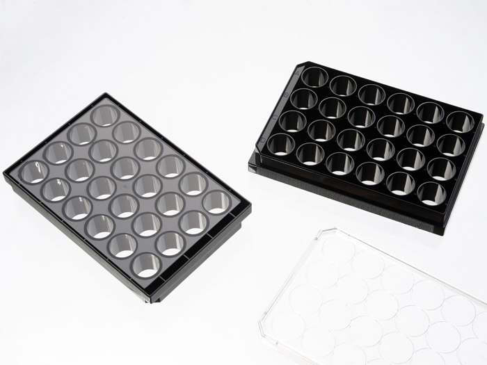
24 Well glass bottom plate with high performance #1.5 cover glass


24 well glass bottom plate with high performance #1.5 cover glass (0.170±0.005mm). Black frame, with lid. Individually packed. Designed for high resolution imaging such as confocal microscopy.
489 cases in stock
** Non-US users please sign in or get a quote to view the proper price for your country.**
Features:
- Suitable for long term tissue culture
- Manufactured in a class 100,000 clean room
- Frame made from virgin polystyrene.
- German high quality cover glass of superior optical quality, glass thickness is 0.170±0.005mm
- A USP class VI adhesive is used to assemble the cover glass and the plate.
- Sterilized by Gamma radiation.
- Conforms to ANSI/SBS 1-2004 standards
Suitable for:
- Differential Interference Contrast (DIC)
- Widefield Fluorescence
- Confocal Microscopy
- Two-Photon and Multiphoton Microscopy
- Fluorescence Recovery After Photobleaching (FRAP)
- Förster Resonance Energy Transfer (FRET)
- Fluorescence Lifetime Imaging Microscopy (FLIM)
- Total Internal Reflection Fluorescence (TIRF)
- Super-Resolution Microscopy
Recommended for:
- Confocal Microscopy
- Super-Resolution Microscopy
Technical specifications
» View technical specification of different coverslips.
| Frame color | black |
|---|---|
| Coverslip | #1.5 high performance cover glass (0.170±0.005mm) |
| Length | 127.50 mm |
| Width | 85.25 mm |
| Height | 20 mm |
| Height with lid | 22.5 mm |
| Stacking height (with lid) | 19.5 mm |
| Bottom height | 0.83 mm (bottom of coverslip to plate bottom) |
| Bottom height tolerance | ±50μm (whole plate) |
| Well to well center distance | 19.3 mm |
| Well bottom area | 190 mm2 |
| Maximum volume | 3.6 ml |
| Temperature Range | -20°C to 50°C |
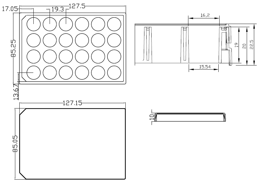
Dimension diagram (units in mm)

Cited Publications before 2019 (20)
-
Peroxisome-derived lipids regulate adipose thermogenesis by mediating cold-induced mitochondrial fission
Hongsuk Park, et al., J. of Clinical Inves. December 4, 2018
Quote: "BAT SVF cells stably expressing Mito-roGFP were seeded in 24-well plates with high performance #1.5 cover glass bottoms (Cellvis #P24-1.5H-N)," -
In vitro characterization of the human segmentation clock
Margarete Diaz-Cuadros, et al., BioRxiv November 04, 2018.
Quote: "except cells were seeded on 35 mm matrigel-coated glass-bottom dishes (MatTek cat. no. P35G-1.5-20-C) or 24 well glass-bottom plates (In Vitro Scientific cat. no. P24-1.5H-N). DMEM/F12 without phenol red was used to reduce background fluorescence (Gibco cat. no. 21041025 )." -
Bright split red fluorescent proteins with enhanced complementation efficiency for the tagging of endogenous proteins and visualization of synapses
S Feng, et al., BioRxiv, October 25, 2018.
Quote: "24-well plates with #1.5 glass coverslip bottoms (In Vitro Scientific) and transfected ~24 hours before measurement using GenJet transfection reagent (SignaGen Laboratories) according to the manufacturer's instructions" -
Investigating LINC complex protein homo-oligomerization in the nuclear envelopes of living cells using fluorescence fluctuation spectroscopy
Jared Hennen, et al., Methods in Molecular Biology, The LINC Complex pp 121-135
Quote: "The FFS experimental setup used in this work. 2.2 Samples and Microscope Slide. 1. 24-well glass-bottom slide with #1.5H cover glass (In Vitro Scientific, Sunnyvale, CA)" -
Controllable protein phase separation and modular recruitment to form responsive membraneless organelles
Benjamin S. Schuster, et al., Nature Communications volume 9, Article number: 2985 (2018)
Quote: "HEK293, HeLa, and U2OS cells were plated at a density of 75 000 cells/well in 0.5 mL Dulbecco’s modified Eagle’s medium (supplemented with 10% fetal bovine serum, glutamine, and pen/strep) in a 24-well glass-bottom plate (#1.5 cover glass; Cellvis) and incubated using standard tissue culture methods at 37 °C with 5% CO2 and saturating humidity" -
Fluorescence fluctuation spectroscopy reveals differential SUN protein oligomerization in living cells
Jared Hennen, et al., Molecular Biology of the CellVol. 29, No. 9
Quote: "Twelve hours prior to their transfection, U2OS cells were grown in 24-well plates with #1.5 glass coverslip bottoms or 35 mm dishes with #1.5 glass coverslip bottoms from In Vitro Scientific" -
ONC201 kills breast cancer cells in vitro by targeting mitochondria
Yoshimi Endo Greer, et al., Oncotarget. 2018 Apr 6; 9(26): 18454–18479.
Quote: "Time-lapse live cell imaging: MB231 cells were seeded (60,000 cells/well) on a 24 well glass bottom plates (Part#P24-1.5H-N, CellVis, Mountain View, CA, USA) with RPMI1640 growth medium." -
Rapid, topology-based particle tracking for high-resolution measurements of large complex 3D motion fields
Mohak Patel, et al., Nature: Scientific Reportsvolume 8, Article number: 5581 (2018)
Quote: "Soft polyacrylamide (PA) gels were prepared as previously described by Toyjanova et al.17. in 24-well glass bottom plates (Cellvis, P24-1.5H-N)." -
Endoplasmic reticulum–resident protein 57 (ERp57) oxidatively inactivates human transglutaminase 2
Michael C. Yi, et al., JBC, February 23, 2018
Quote: "HUVECs were grown to 70% confluence in a 24-well glass bottom plate (catalog no. P24–1.5H-N, CellVis)." -
Two water-soluble two-photon fluorescence probes for ratiometric imaging endogenous SO2 derivatives in mitochondria
M Zhao, et al., Sensors and Actuators B: Chemical Volume 255, Part 2, February 2018, Pages 1228-1237
Quote: "For live cell confocal laser scanning microscopy experiment, HepG2 cells were seeded in 24-well glass bottom plate (In Vitro Scientific, P24-1.5H N) at density of 10,000, and incubated for 72–96 h at 37 °C in 95% air 5% CO 2 in order to allow the cells to reach ∼90"
View all publications citing "24 Well glass bottom plate with high performance #1.5 cover glass".


