


24 Well Plate with #1.5 glass-like polymer coverslip bottom, tissue culture treated for better cell attachment than cover glass
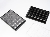
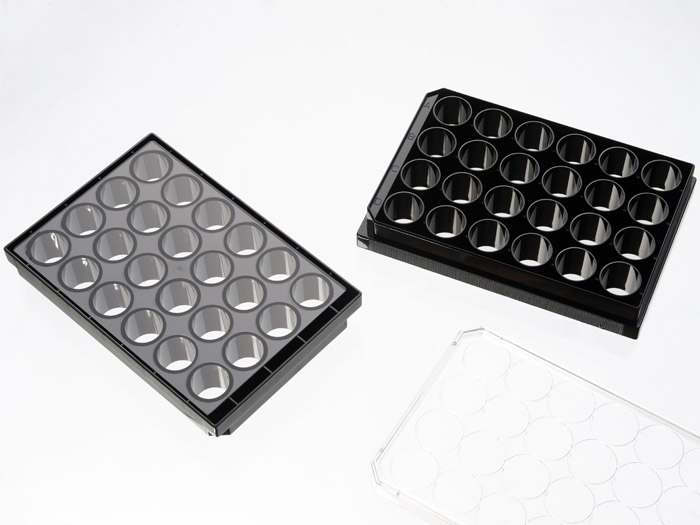
24 well plate with tissue culture treated #1.5 glass-like polymer coverslip (0.175±0.010mm). Tissue culture treated. Black frame with lid. Designed for high resolution imaging such as confocal microscopy.
Recommended for applications where better cell attachment is desired. Not suitable for total internal reflection fluorescence (TIRF). May not be suitable for applications such as whole plate auto-scan, for which whole plate flatness is critical.
114 cases in stock
** Non-US users please sign in or get a quote to view the proper price for your country.**
Features:
- Suitable for long term tissue culture
- Frame made from virgin polystyrene.
- Better cell attachment with #1.5 glass-like polymer coverslip (0.175±0.010mm)
- Manufactured in a class 100,000 clean room
- A USP class VI adhesive is used to assemble the coverslip and the plate.
- Sterilized by Gamma radiation.
- Conforms to ANSI/SBS 1-2004 standards
Suitable for:
- Differential Interference Contrast (DIC)
- Widefield Fluorescence
- Confocal Microscopy
- Two-Photon and Multiphoton Microscopy
- Fluorescence Recovery After Photobleaching (FRAP)
- Förster Resonance Energy Transfer (FRET)
- Fluorescence Lifetime Imaging Microscopy (FLIM)
- Super-Resolution Microscopy
Recommended for:
- Confocal Microscopy
Not suitable for:
- Total Internal Reflection Fluorescence (TIRF)
Technical specifications
» View technical specification of different coverslips.
| Frame color | black |
|---|---|
| Coverslip | #1.5 glass-like polymer (0.175±0.010mm) |
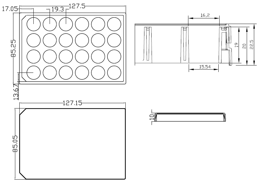
| Length | 127.50 mm |
| Width | 85.25 mm |
| Height | 20 mm |
| Height with lid | 22.5 mm |
| Bottom height | 0.83 mm (bottom of coverslip to plate bottom) |
| Stacking height (with lid) | 19.5 mm |
| Well to well center distance | 19.3 mm |
| Well bottom area | 190 mm2 |
| Maximum volume | 3.6 ml |
| Temperature Range | -20°C to 50°C |
Dimension diagram (units in mm)
Open Source Publications Citing This Product
-
Uncovering cancer dependencies in peptide-interacting protein pockets
M Örd, L Kogan, D Bradburn, M Leiser, M Kõivomägi - … 2026
… HCT116 cells transduced with HES1 WRPW-GFP expression constructs were grown to 30% confluency in a Cellvis P24-1.5P 24-well plate with polymer coverslip base (… -
Benchmarking Machine Learning and Automated Image Analysis for Organelle Quantification
C Daul, P Tournier, SJ Habib - Chemical & Biomedical Imaging 2026
… For all experiments, the cells were seeded at a density of 2.6 × 10 3 cells/cm 2 in Cellvis 24-well imaging bottom plates (P24-1.5P) and cultured for 48 h prior to imaging, … -
Toward an all-in-one recombinant adeno-associated virus vector for functionally ablating the prion gene using CRISPR-Cas technology
C Verkuyl, A Belotserkovsky, T Zerbes, D Williams - Plos one 2025
… 1x10⁵ cells were seeded on a 24 well glass imaging plate (catalog number P24-1.5P, Cellvis, Mountain View, CA, USA) and proliferated to ~70–80% confluence prior to … -
Mitotic DNA repair by TMEJ suppresses replication stress-induced nuclear envelope reassembly defect
G Ye, Y He, Y Zhang, D Li, F Liu, Y Li, Q Ge - Nature … 2025
Replication stress (RS), if not effectively and timely addressed, could result in DNA damage in mitosis. However, the relationship between RS and other mitotic events, … -
Uncovering cancer dependencies in peptide-interacting protein pockets
M Örd, L Kogan, D Bradburn, M Leiser, M Kõivomägi - europepmc.org
… (which was not certified by peer review) is the author/funder, who has granted bioRxiv a license to display the preprint in perpetuity. It is made The copyright holder for this …