96 Well glass bottom plate with #1 cover glass
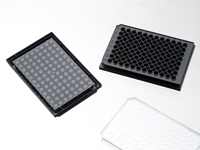

96 well glass bottom plate. Black polystyrene frame with #1 glass(0.13-0.16mm), with lid, Individually packed. Designed for high resolution imaging such as confocal microscopy.
27 cases in stock
** Non-US users please sign in or get a quote to view the proper price for your country.**
Features:
- Suitable for long term tissue culture
- Manufactured in a class 100,000 clean room
- Frame made from virgin polystyrene.
- German cover glass of superior optical quality
- A USP class VI adhesive is used to assemble the cover glass and the plate.
- Sterilized by Gamma radiation.
- Conforms to ANSI/SBS 1-2004 standards
Suitable for:
- Differential Interference Contrast (DIC)
- Widefield Fluorescence
- Confocal Microscopy
- Two-Photon and Multiphoton Microscopy
- Fluorescence Recovery After Photobleaching (FRAP)
- Förster Resonance Energy Transfer (FRET)
- Fluorescence Lifetime Imaging Microscopy (FLIM)
- Total Internal Reflection Fluorescence (TIRF)
- Super-Resolution Microscopy
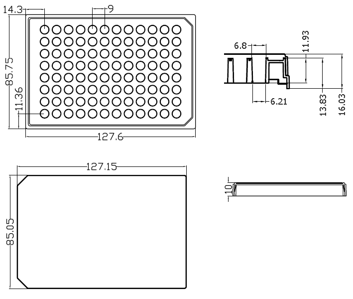
Technical specifications
» View technical specification of different coverslips.
| Frame color | black |
|---|---|
| Coverslip | #1 cover glass (0.13-0.16 mm) |
| Length | 127.60 mm |
| Width | 85.75 mm |
| Height | 14.30 mm |
| Height with lid | 16.5 mm |
| Stacking height (with lid) | 15.5 mm |
| Bottom height | 1.75 mm (bottom of coverslip to plate bottom) |
| Bottom height tolerance | ±50μm (whole plate) |
| Lid dimension | 127 * 85 * 10 mm |
| Well to well center distance | 9.00 mm |
| Well bottom area | 30 mm2 |
| Maximum volume | 0.35 ml |
| Temperature Range | -20°C to 50°C |
Dimension diagram (units in mm)
Cited Publications before 2019 (2)
-
A FRET-Based approach to study the SUMOylation of Serotonin 1A Receptors
S Kaur, et al., Ku Schoolworks, 2018
Quote: "or in 96-well glass bottom plates (Catalog no. #P96-1-N, CellVis, Mountain View, CA, USA) at a seeding density of 15,000 cells per well." -
Metabolic origins of spatial organization in the tumor microenvironment
Carlos Carmona-Fontaine, et al., PNAS
Quote: "Tube Formation Assay. Macrophages alone or cocultured with tumor cell lines were cultured in glass bottom 96-well plates (Cellvis; P96-1-N)"