29 mm Glass bottom dish with 20 mm micro-well #1 cover glass

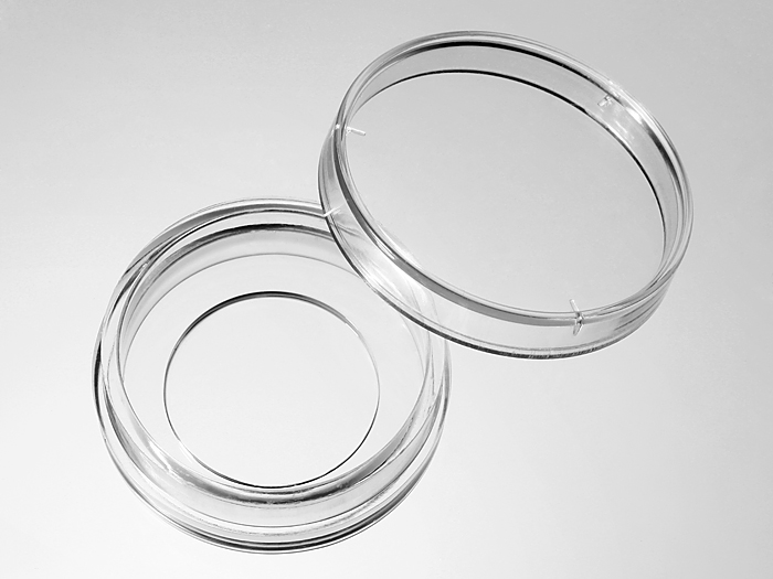
29 mm glass bottom dish, dish size 29mm, well size 20mm, #1 cover glass(0.13-0.16mm). Designed for high resolution imaging such as confocal microscopy.
20 cases in stock
** Non-US users please sign in or get a quote to view the proper price for your country.**
Features:
- Suitable for long term tissue culture
- Manufactured in a class 100,000 clean room
- Dish made from virgin polystyrene, tissue culture treated.
- German cover glass of superior optical quality
- A USP class VI adhesive is used to assemble the cover glass and the dish.
- Packed in easy to open peelable bag
- Sterilized by Gamma radiation.
Suitable for:
- Differential Interference Contrast (DIC)
- Widefield Fluorescence
- Confocal Microscopy
- Two-Photon and Multiphoton Microscopy
- Fluorescence Recovery After Photobleaching (FRAP)
- Förster Resonance Energy Transfer (FRET)
- Fluorescence Lifetime Imaging Microscopy (FLIM)
- Total Internal Reflection Fluorescence (TIRF)
- Super-Resolution Microscopy
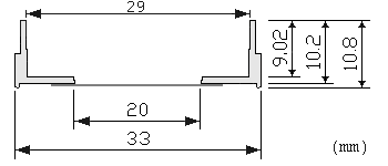
Technical specifications
» View technical specification of different coverslips.
| Coverslip | #1 cover glass (0.13 - 0.16 mm) |
|---|---|
| Temperature Range | -20°C to 50°C |
| Lid diameter (outer) | 34.3 mm |
Dimension diagram (units in mm)
Cited Publications before 2019 (2)
-
A novel probe for phosphatidylinositol 4-phosphate reveals multiple pools beyond the Golgi
Gerald R.V. Hammond, et al., The Journal of Cell Biology, April 2014, Vol. 205 no. 1 113-126
Quote: "Cells were seeded in 29-mm dishes with 20-mm #1 cover glass bottoms (In Vitro Scientific) at appropriate densities so that cells reached 50–100% confluence on the day of imaging" -
Visualizing Escherichia coli Sub-Cellular Structure Using Sparse Deconvolution Spatial Light Interference Tomography
Mustafa Mir, S. Derin Babacan, Michael Bednarz, Minh N. Do, Ido Golding, Gabriel Popescu, Plosone, June 2012, Volume 7, issue 6
Quote: "The cells are then concentrated to an OD of ~0.4 and 2 µl of the culture ispipetted onto a glass bottom dish (In Vitro Scientific D29-20-1-N) and covered bya 1 mm thick agar slab (1.5% Agarose, M9CA media)."