


384 Well glass bottom plate with high performance #1.5 cover glass
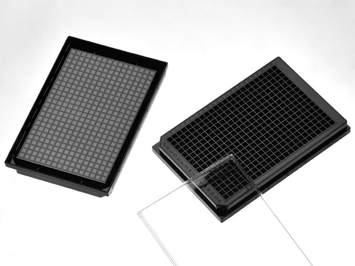
384 well glass bottom plate. Black polystyrene frame with high performance #1.5 cover glass (0.170±0.005mm), with lid, Individually packed. Designed for high resolution imaging such as confocal microscopy.
109 cases in stock
** Non-US users please sign in or get a quote to view the proper price for your country.**
Features:
- Suitable for long term tissue culture
- Manufactured in a class 100,000 clean room
- Frame made from virgin polystyrene.
- German high quality cover glass of superior optical quality, the cover glass has a thickness of 0.170±0.005mm
- A USP class VI adhesive is used to assemble the cover glass and the plate.
- Sterilized by Gamma radiation.
- Conforms to ANSI/SBS 1-2004 standards
Suitable for:
- Differential Interference Contrast (DIC)
- Widefield Fluorescence
- Confocal Microscopy
- Two-Photon and Multiphoton Microscopy
- Fluorescence Recovery After Photobleaching (FRAP)
- Förster Resonance Energy Transfer (FRET)
- Fluorescence Lifetime Imaging Microscopy (FLIM)
- Total Internal Reflection Fluorescence (TIRF)
- Super-Resolution Microscopy
Recommended for:
- Confocal Microscopy
- Super-Resolution Microscopy
Technical specifications
» View technical specification of different coverslips.
| Frame color | black |
|---|---|
| Coverslip | #1.5 high performance cover glass (0.170±0.005mm) |
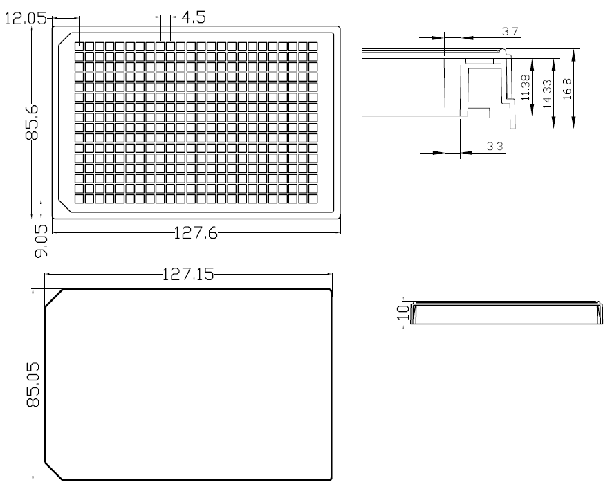
| Length | 127.60 mm |
| Width | 85.60 mm |
| Height | 14.33 mm |
| Height with lid | 16.80 mm |
| Bottom height | 2.78 mm (bottom of coverslip to plate bottom) |
| Bottom height tolerance | ±50μm (whole plate) |
| Well to well center distance | 4.50 mm |
| Well bottom area | 10.89 mm2 |
| Maximum volume | 0.12 ml |
| Temperature Range | -20°C to 50°C |
Dimension diagram (units in mm)
Open Source Publications Citing This Product
-
Advanced Open-source Experimental-Design Tools for Microplate-Based Assays with Acoustic Liquid Handling
VM Kattunga, SA Wrobel, CA Lerner, VM Derycz - bioRxiv 2026
… All other ADE happened into 384-well glass-bottomed plates (Cellvis #P384-1.5HN). For manual culturing of dispersed human pancreatic β-cells, glass-bottomed 96-well … -
Opposing roles of serine and charge in IDR condensate miscibility
G Pei, X Wang, X Quan, D Geng, Z Chen, W Xu - Nature Chemical … 2026
Numerous biomolecular condensates coexist within cells, yet the factors governing their miscibility remain poorly understood. Here, by examining 28 intrinsically … -
Efficient Generation of Functional TCRαβ+ Cytotoxic T Cells from hiPSCs via Small-Molecule Modulation
C Kubaczka, N Kambli, R Windisch, K Yu, Y Zhao - … 2026
… Rows AO of a glass bottom 384-well plate (Cellvis, #P384–1.5HN) were coated with DLL4 (Thermo Fisher) and VCAM1 by adding 25 μl/well with a 300 μl 12-channel … -
A calcium imaging pipeline to detect and quantify compound-specific effects in human and mouse astrocytes and astrocyte-neuron cocultures
J Krohn, L Breuer, S Wegmann, C Dean - bioRxiv 2026
… effects in human and mouse astrocytes and astrocyte-neuron cocultures | bioRxiv … , 200 µL culture medium; 384-well (Cellvis, P384-1.5HN): ∼120,000 cells/cm², 100 µL … -
The Spatiotemporal Proteome Landscape of Aging: Structural determinants of age-sensitive proteome remodeling
S Yoo, L Vannur, L Li, C Young, Q Liu, ZT Wen - bioRxiv 2026
… Cells were imaged in 384-well glass-bottom plates (Cellvis, P384-1.5HN, USA). Images were 815 acquired on a Nikon CSU-W1 SoRa spinning-disk confocal microscope … -
Single-Cell Spatial Proteomics Uncovers Molecular Interconnectivity among Hallmarks of Aging
S Yoo, C Young, L Li, L Vannur, J Zhuang, F Zheng - bioRxiv 2026
Aging is accompanied by conserved hallmarks including genomic instability, epigenetic alterations, loss of proteostasis, and mitochondrial dysfunction, but how these … -
Revealing site-specific molecular features mediating contacts of heterochromatin protein 1 α (HP1α) and the interactions with chromatin and DNA in LLPS …
Z Zhai, A Soman, J Kuang, J Guo, Y Chen - Communications … 2025
… The 384-Well Microscopy Plates (Cellvis, #P384-1.5HN) were washed and then pre-treated with 1 M NaOH and 5 K mPEG-silane as described in one of our recent studies… -
Liquid-liquid phase separation of ATXN2L enhances mRNA translation in hepatocellular carcinoma
S Wang, P Guo, W Ma, H Huang, Q Zhang - Cell Reports 2025
… The samples were then transferred into a 386-well plate (P384-1.5HN, CellVis, Mountain View, USA) for observation. Imaging of the phase-separated structures was … -
Explainable AI to unveil cellular autophagy dynamics
O Presacan, M Hernández Mesa, AC Aldea - Plos one 2025
… Cells were imaged in a 384-well glass-bottom plate (Cellvis, P384-1.5HN) pre-coated with 0.5 mg/mL concanavalin A (Sigma, L7647). Live-cell time-lapse imaging was … -
Fluorogenic RNA Aptamer-Based Activity Quantification of T7 RNA Polymerase
K Mimouni, W Qiu, H Zhou - Biochemistry
… Once prepared, Broccoli IVT reactions were transferred to a black-edge and clear-bottom 384-well plate (Cellvis P384-1.5HN) and monitored using a Neo2 plate reader (… -
2050-084X
L Adèle - eLife
… P384-1.5HN) were washed with PBS and then permeabilized with 0.1% Triton X-100 (Sigma, Cat. No. T9284-500ML) solution in PBS for 15 minutes and again washed …