


6 Well glass bottom plate with high performance #1.5 cover glass
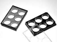
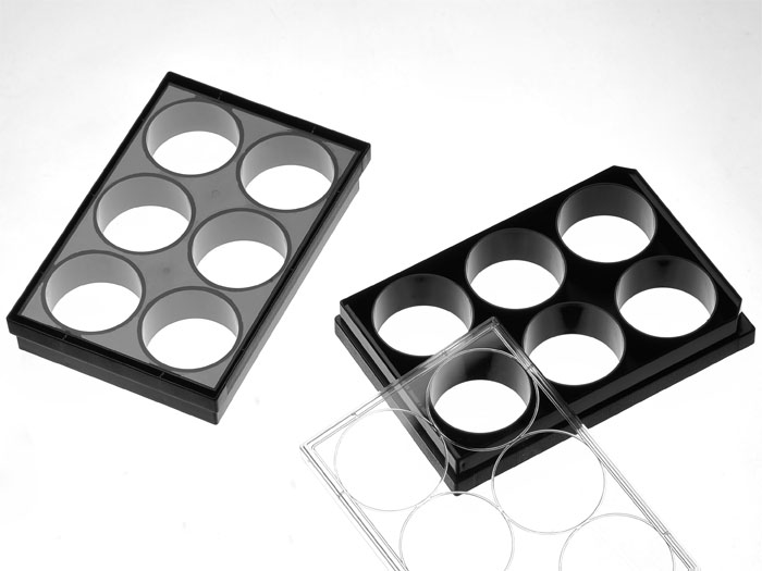
6 well glass bottom plate with high performance #1.5 cover glass (0.170±0.005mm). Black frame, with lid. Individually packed. Designed for high resolution imaging such as confocal microscopy.
134 cases in stock
** Non-US users please sign in or get a quote to view the proper price for your country.**
Features:
- Suitable for long term tissue culture
- Manufactured in a class 100,000 clean room
- Frame made from virgin polystyrene
- German high quality cover glass of superior optical quality, glass thickness is 0.170±0.005mm
- A USP class VI adhesive is used to assemble the cover glass and the plate.
- Sterilized by Gamma radiation.
- Conforms to ANSI/SBS 1-2004 standards
Suitable for:
- Differential Interference Contrast (DIC)
- Widefield Fluorescence
- Confocal Microscopy
- Two-Photon and Multiphoton Microscopy
- Fluorescence Recovery After Photobleaching (FRAP)
- Förster Resonance Energy Transfer (FRET)
- Fluorescence Lifetime Imaging Microscopy (FLIM)
- Total Internal Reflection Fluorescence (TIRF)
- Super-Resolution Microscopy
Recommended for:
- Confocal Microscopy
- Super-Resolution Microscopy
Technical specifications
» View technical specification of different coverslips.
| Frame color | black |
|---|---|
| Coverslip | #1.5 high performance cover glass (0.170±0.005mm) |
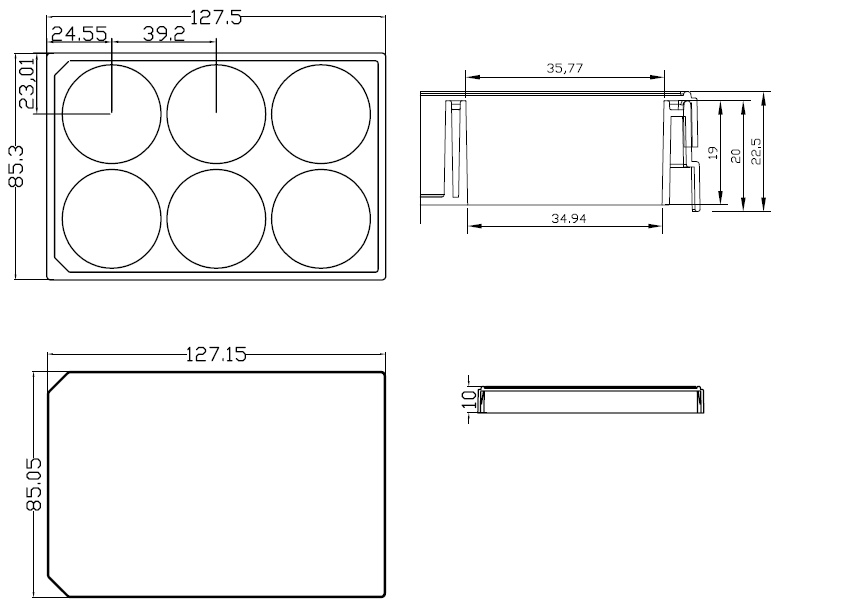
| Bottom length | 127.50 mm |
| Bottom width | 85.30 mm |
| Height | 20 mm |
| Height with lid | 22.5 mm |
| Stacking height | 19.5 mm |
| Bottom height | 0.83 mm (bottom of coverslip to plate bottom) |
| Bottom height tolerance | ±50μm (whole plate) |
| Well bottom area | 958 mm2 |
| Maximum volume | 18 ml |
| Temperature Range | -20°C to 50°C |
Dimension diagram (units in mm)
Open Source Publications Citing This Product
-
Understanding the Control Logic of Subcellular Self-Organization: From Ciliate Morphology to Biomolecular Condensates
ZM Xu - 2026 2026
Biological systems self-organize internal components into functional structures across scales through the dynamic interplay between intracellular regulatory networks and … -
Serotonin Receptor Signaling Promotes Amoeboid Migration of Melanoma Cells Through cAMP/PKA Activation
AG Abrenica, S Kuang, N Kar, JS Logue - bioRxiv 2026
… CC-BY 4.0 International license available under a (which was not certified by peer review) is the author/funder, who has granted bioRxiv a license to display the preprint in … -
Systematic functional genetic analysis of cell-substrate adhesion
K Manzer, KC Su, MM Di Bernardo, HR Keys, R Walton - bioRxiv 2026
… KO HeLa cell library was induced with 1 μg/mL doxycycline hyclate for 78 hours and seeded at a density of 350k cells/well in 6-well glassbottom plates (Cellvis, P06-1.5HN… -
Revealing the spatiotemporal dynamics of methionine metabolism with a genetically encoded single-fluorophore biosensor
KA Cohen, A Jhawar, G Garcia, N Goni, M Chen - bioRxiv 2026
… Cells were seeded and grown to 70-80% confluency in 6-well glass bottom dishes (Cellvis Cat #P06-1.5HN) prior to treatment with small molecules. Following treatment, … -
Leptin drives osteoarthritis pain through sensory neuron reprogramming independent of cartilage signaling
HD Welhaven, EK Truchan, KL Lenz, BA Andoko - bioRxiv 2026
… The cells were then plated on 6 well glass bottom plates (P06-1.5HN) precoated with 0.1mg/mL of poly-D-lysine (Sigma-Aldrich, P7405-5MG). The cultures were placed in … -
Scalable multimodal mapping of macrophage regulatory architecture by integrating optical and transcriptomic pooled screens
T Kudo, R Lopez, AM Meireles, A Rios, P Coelho - bioRxiv 2026
Understanding how genetic perturbations reshape cellular states requires measuring diverse phenotypic modalities at scale. Here, we present PerturbPair, a platform that … -
A Far-Red FRET biosensor for AMPK enables multiplexed imaging of single-cell bioenergetic homeostasis.
NL DeCuzzi, M Hardy, MB Cabel, J Hu - Biorxiv: the Preprint … 2026
… (which was not certified by peer review) is the author/funder, who has granted bioRxiv a license to display the preprint in perpetuity. It is made The copyright holder for this … -
A unified photosensitizer platform for in situ DNA-, RNA-, and protein-directed proximity labeling
EB Biletch, CP Herlihy, L Li, M Krebs, CJ Kelly - … 2026
… For each ISH-POCA experiment, 300,000 cells were seeded into each well of a glass-bottom 6-well plate (Cellvis P06-1.5HN) to culture for 24 hours to reach 750,000 - 1 … -
The one-week automated genome-wide optical pooled screen
B Kirby, M Di Bernardo, IM Cheeseman, PC Blainey - bioRxiv 2026
… a 24-hour incubation, cells were washed once with 1X PBS, passaged with TrypLE (Thermo Fisher Scientific 12604013), and seeded in two six-well plates (P06-1.5HN) at … -
A defined 2D system for generating and expanding human basal radial glia from iPSCs
A Artioli, M Gasparotto, AC Rossetti, Y Hass - bioRxiv 2026
… For live imaging of IST events, bRG cells were plated onto 6-well glass bottom plates (IBL P06-1.5HN), GT-coated and imaged on a Nikon TI2-E inverted video-microscope … -
Quantitative live imaging of junctional neural tube formation
J Wang - 2026 2026
… The embryos were then mounted with mounting media (Sigma-Aldrich, F4680-25ML) on a six-well glass bottom plate (Cellvis, P061.5HN) for imaging or stored at 4oC in … -
Long-range actin-driven endosymbiont mobility in an animal-algal symbiosis
G Zhong, G Toullec, PH Jouneau - Available at SSRN … 2026
… For Waminoa sp., worms were placed in glass bottom dishes (Cellvis, P06-1.5HN), and allowed 384 to settle on the microscope for 30 minutes up to a few hours without … -
Cell. Author manuscript; available in PMC 2020 October 17. Published in final edited form as: Cell. 2019 October 17; 179 (3): 787–799. e17. doi: 10.1016/j. cell …
D Feldman, A Singh, JL Schmid-Burgk, RJ Carlson - dspace.mit.edu
polymerase, 0.5 U/μL Ampligase and 50 nM dNTPs) for 5 minutes at 37C and 90 minutes at 45C, and then washed 2 times with PBS-T. Circularized padlocks were … -
2050-084X
KY Tony, W Luke, J Laura, ES John - eLife
… Unless otherwise specified, black glass-bottom 6-well plates (P06-1.5HN, Cellvis) were used for microplate imaging. Prior to imaging, the FLS media was replaced with …